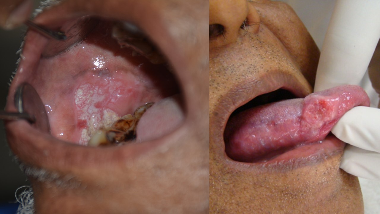
Oral cancer is a serious health challenge to the most of the developing countries. As per various studies the data suggests that in India, around 77,000 new cases and 52,000 deaths are reported annually, which is approximately one-fourth of global incidences. The increasing cases of oral cancer are the significant issue for a community health as it is one of the common types of cancers in India.
As compared to the other countries in west, the concern of oral cancer is significantly higher in India as about 70% of the cases are reported in the advanced stages. It usually happens that because of its detection in the late phase, the chances of cure are very low, almost negative; leaving five-year survival rates around 20% only.
At CIMS, our oral and maxillofacial consultant carry out a detailed physical examination. The latest technology, for imaging, or microscopic evaluation can be used eventually to hasten the treatment planning.
A specialist’s experience in visually analyzing cancers is the leading factor, although scientific back up should be used to aid their view and the diagnosis extends beyond what can be seen on your face, or within your mouth. Hence the needful procedures such as various types of biopsies are carried out to identify any sinister lesions.
The evolution of dentistry in recent decades has been rapid and remarkable. Every moment a new technique is described, mastered, and popularized. In the not-too-distant past, dental treatment consisted of seeking pain relief; often extracting the tooth was considered the most effective treatment.
Contemporary dentistry seeks the preservation and restoration of teeth, periodontal tissues, and peri-implant tissues, with an appropriate relationship between the arches. The treatment philosophy should focus on the restoration of dentofacial function and esthetics to provide or restore the patient’s physical, mental, and social well-being, improving their quality of life.
Often dissociated, esthetics and function are integral parts of the same system. They must act synergistically to provide greater predictability and longevity to dental treatments.
In the field of esthetics, many subjective components are linked to ethnicity, belief, culture, age, and individuality. However, there are rules and parameters that, when observed, become a good starting point for the dentist to develop a clinical and digital plan of the rehabilitating treatments.
Care will be managed by the surgical team at CIMS. They are experienced oral & maxillofacial surgeons and can call on our multidisciplinary team, allowing us to offer wide support.
Hence, each member of our staff understands this and is dedicated to seeing our patients enjoy their lives again. Whether we are treating an isolated issue, or a multi faceted condition, the same ethos applies.
Exceptional care and the facilities you need will be there to support you. You are welcome to talk to our friendly team at any time.

Basal cell carcinoma is a type of skin cancer. Basal cell carcinoma begins in the basal cells — a type of cell within the skin that produces new skin cells as old ones die off.
Basal cell carcinoma often appears as a slightly transparent bump on the skin, though it can take other forms. Basal cell carcinoma occurs most often on areas of the skin that are exposed to the sun, such as your head and neck.
Most basal cell carcinomas are thought to be caused by long-term exposure to ultraviolet (UV) radiation from sunlight. Avoiding the sun and using sunscreen may help protect against basal cell carcinoma.
Basal cell carcinoma appears as a change in the skin, such as a growth or a sore that won’t heal. These changes in the skin (lesions) usually have one of the following characteristics:
A shiny, skin-colored bump that’s translucent, meaning you can see a bit through the surface. The bump can look pearly white or pink on white skin. On brown and Black skin, the bump often looks brown or glossy black. Tiny blood vessels might be visible, though they may be difficult to see on brown and Black skin. The bump may bleed and scab over.
A brown, black or blue lesion — or a lesion with dark spots — with a slightly raised, translucent border.
A flat, scaly patch with a raised edge. Over time, these patches can grow quite large.
A white, waxy, scar-like lesion without a clearly defined border.
Make an appointment with us if you observe changes in the appearance of your skin, such as any new growth or any change in a previous growth or any recurring sore.
Melanoma, which means “black tumor,” is the most dangerous type of skin cancer. It grows quickly and has the ability to spread to any organ.
Melanoma comes from skin cells called melanocytes. These cells produce melanin, the dark pigment that gives skin its color. Most melanomas are black or brown in color, but some are pink, red, purple or skin-colored. About 30% of melanomas begin in existing moles, but the rest start in normal skin. This makes it especially important to pay attention to changes in your skin because the majority of melanomas don’t start as moles.
However, how many moles you have may help predict your skin’s risk for developing melanoma. Knowing your risk can help you be extra vigilant in watching changes in your skin and seeking skin examinations since melanomas have a 99% cure rate if caught in the earliest stages. Early detection is important because treatment success is directly related to the depth of the cancerous growth.
Sarcoma is a type of cancer that can occur in various locations in your body. It is a general term for a broad group of cancers that begin in the bones and in the soft (also called connective) tissues (soft tissue sarcoma). According to various studies there are more than 70 types of sarcoma. Treatment for sarcoma varies depending on sarcoma type, location and other factors.